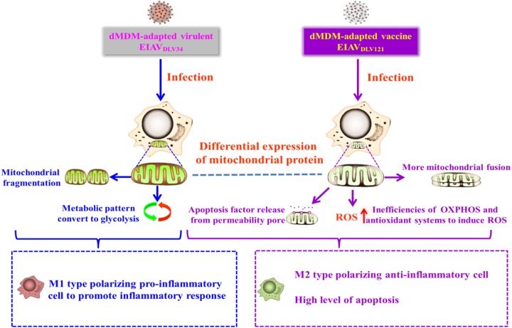
FIG 9.
Schematic diagrams demonstrating the role of mitochondria in the regulation of the intracellular environment to produce an inflammatory response or apoptosis after EIAVDLV34 or EIAVDLV121 infection. (Left) After EIAVDLV34 infection, alteration of the expression pattern of mitochondrial proteins leads to the conversion of the metabolic pattern to glycolysis and mitochondrial fragmentation, which facilitate the transformation of eMDMs to M1-type-polarizing proinflammatory cells. Meanwhile, the cells keep normal apoptosis and ROS levels. Ultimately, the inflammatory response occurs. (Right) After EIAVDLV121 infection, alteration of the expression pattern of mitochondrial proteins leads to inefficiencies of OXPHOS and antioxidant systems, dissipation of the Δψm, more mitochondrial fusion, and inhibition of glycolysis, which facilitate increases of apoptosis and ROS levels and transformation of eMDMs to M2-type-polarizing anti-inflammatory cells. Ultimately, inflammation keeps balance and additional apoptosis is induced.

