FIG 2.
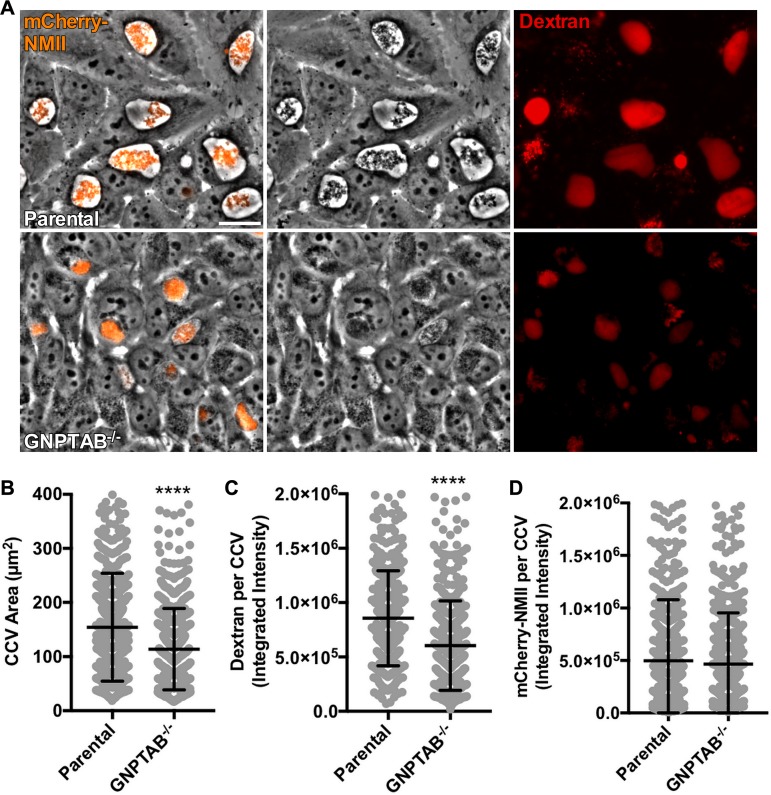
GNPTAB−/− HeLa cells support C. burnetii growth, but CCVs are abnormal. (A to C) C. burnetii growing in GNPTAB−/− cells generates dark, condensed CCVs with decreased dextran fluorescence. Cells were infected with mCherry-NMII for 3 days, incubated for 18 h with Alexa Fluor 647 dextran, and imaged live. Images were analyzed for CCV area and dextran fluorescence intensity. (D) The growth of C. burnetii per vacuole is normal in GNPTAB−/− cells. Images from panel A were analyzed for mCherry-NMII fluorescence intensity per CCV. Graphs represent the means ± SD of ≥150 cells from 3 independent experiments. Statistical significance was determined by Student's t test (****, P < 0.0001). Scale bar, 10 μm.

