FIG 4.
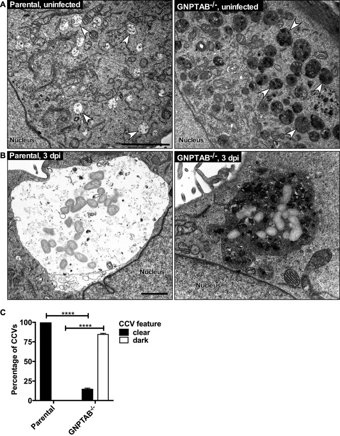
CCVs in GNPTAB−/− HeLa cells accumulate electron-dense material. (A) By TEM, uninfected GNPTAB−/− cells contain abundant inclusions, observed as enlarged, dark endocytic vesicles. Arrows point to representative late endocytic vesicles. (B and C) CCVs in parental cells are clear and spacious, whereas the majority of CCVs in GNPTAB−/− cells are dense and filled with material. TEM images are of cells at 3 dpi. Images were analyzed for percentage of clear versus dark vacuoles. Magnification of TEM images was at 11,000×. Graphs represent the means ± SD of ≥60 cells from 2 independent experiments. Statistical significance determined by Student's t test (****, P < 0.0001). Scale bar, 1 μm.

