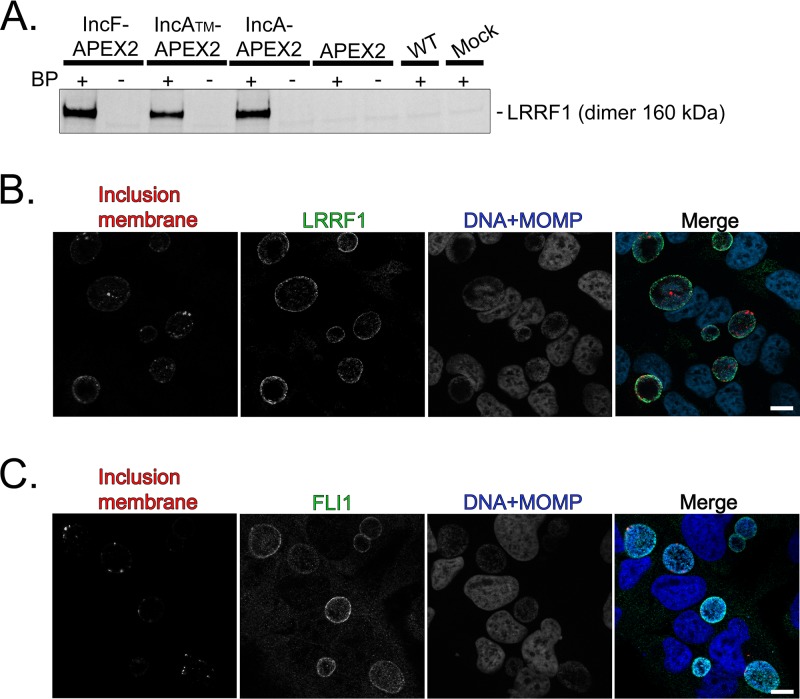
FIG 5.
Confirmation of LRRF1 biotinylation by Inc-APEX2 proteins and localization of LRRF1 and FLII to the chlamydial inclusion. (A) Western blotting confirmation of LRRF1 in the eluates from streptavidin affinity-purified biotinylated lysate from the C. trachomatis L2 IncF-APEX2, IncATM-APEX2, and IncA-APEX2 transformants at 24 hpi (BP, biotin-phenol). (B) Confirmation of LRRF1 colocalization with the inclusion of C. trachomatis L2 wild-type-infected HeLa cells. Cells were fixed at 24 hpi in 4% paraformaldehyde, permeabilized with 0.5% Triton X-100, and then stained for indirect immunofluorescence to visualize the inclusion membrane (CT223; red), LRRF1 (green), and DNA and chlamydiae (DRAQ5 and MOMP; blue). (C) Confirmation of FLII colocalization with the inclusion of C. trachomatis L2 wild-type-infected HeLa cells. Cells were fixed at 24 hpi in 4% paraformaldehyde, permeabilized with 0.5% Triton X-100, amd then stained for indirect immunofluorescence to visualize the inclusion membrane (CT223; red), FLII (green), and DNA and chlamydiae (DAPI and MOMP; blue). Coverslips were imaged using a Zeiss ApoTome.2 fluorescence microscope at ×100 magnification. Bars = 10 μm. See Fig. S4 in the supplemental material.

