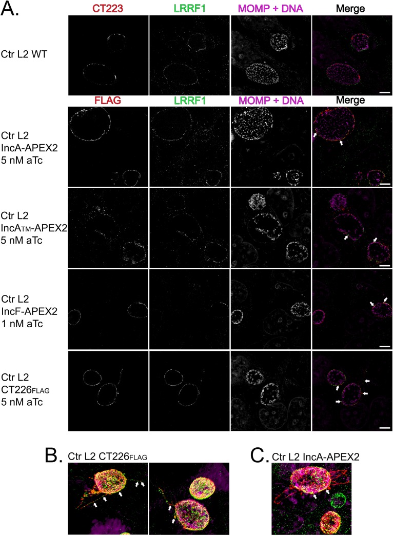
FIG 9.
Assessment of LRRF1 colocalization with chlamydial Incs in C. trachomatis L2-infected HeLa cells using superresolution microscopy. (A) HeLa cells seeded on glass coverslips were infected with C. trachomatis L2 Inc-APEX2 transformants or CT226FLAG transformants and induced for expression at 20 hpi (IncF-APEX2 was induced with 1 nM aTc; 5 nM aTc was used for all other transformants). At 24 hpi, the coverslips were fixed with ice-cold methanol and stained for immunofluorescence to visualize construct expression (FLAG) or CT223 (red), LRRF1 (green), and chlamydiae and DNA (DRAQ5 and MOMP; pink). Coverslips were imaged by structural illumination microscopy (SIM) with a Zeiss Elyra superresolution microscope at a ×63 magnification with a ×2 zoom. Bars = 5 μm. (B) SIM 3D snapshot of C. trachomatis L2 CT226FLAG-infected HeLa cells with CT226FLAG- and LRRF1-positive fibers. (C) SIM 3D snapshot of C. trachomatis L2 IncA-APEX2-infected HeLa cells with IncA fibers. Arrows indicate colocalization between the indicated expressed construct and LRRF1.

