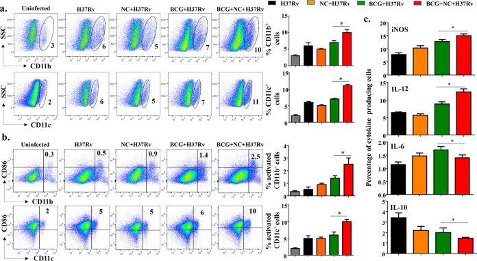
FIG 3.
Nanocurcumin activates APCs in lungs of BCG-vaccinated mice. Mice were treated as described in the legend to Fig. 2a. At 75 days postinfection, lungs were harvested, and single-cell suspensions were made and cultured overnight, followed by staining and flow cytometry. Primarily monocytes were gated based on forward scatter versus side scatter (SSC), and monocytes and other cell types were identified by specific antibodies. (a) Pseudocolor plot and bar diagrams of CD11b+ and CD11c+ APCs in lungs. (b) Activation status of APCs. Pseudocolor plots and bar diagrams of CD11b+ CD86+ and CD11c+ CD86+ APCs in lungs are shown. (c) Bar diagrams of IL-12-, IL-10-, IL-6-, and iNOS-producing APCs. All data are representative of results from 3 independent experiments with 5 mice from each experimental group at each time point. All values are represented as means ± SD. Statistical analyses were done by ANOVA with Tukey’s post hoc analysis. * denotes a P value of ≤0.05. Experimental groups are (i) uninfected, (ii) H37Rv, (iii) nanocurcumin (NC) plus H37Rv, (iv) BCG plus H37Rv, and (v) BCG plus nanocurcumin and H37Rv.

