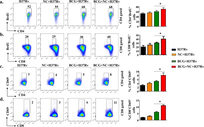
FIG 4.
Nanocurcumin treatment increases the proliferation and activation of antigen-specific CD4+ and CD8+ T cells in BCG-immunized animals. Mice were treated as described in the legend to Fig. 2a. At 72 days postinfection, mice were injected with BrdU. Seventy-two hours after injection of BrdU, spleens were harvested, and single-cell suspensions were made. Cells were cultured overnight with M. tuberculosis lysate (CSA) stimulation to assess antigen-specific immune responses. These cells were stained with anti-CD3, -CD4, -CD8, -CD69, and -BrdU antibodies, followed by flow cytometry. For FACS analysis, we first isolated the CD3 and then the CD4 or CD8 populations, and within those populations, we then gated BrdU+ and CD69+ populations. (a) CD4 T cell proliferative status, as shown by pseudocolor and bar diagrams of CD4+ BrdU+ T cells. (b) CD8 T cell proliferative status, as shown by pseudocolor plots and bar diagrams of CD8+ BrdU+ T cells. (c) CD4 T cell activation status, as shown by pseudocolor plots and bar diagrams of CD4+ CD69+ T cells. (d) CD8 T cell activation status, as shown by pseudocolor plots and bar diagrams of CD8+ CD69+ T cells. All data are representative of results from 3 independent experiments with 5 mice from each experimental group at each time point. All values are represented as means ± SD. Statistical analyses were done by ANOVA with Tukey’s post hoc analysis. * denotes a P value of ≤0.05. Experimental groups are (i) H37Rv, (ii) nanocurcumin (NC) plus H37Rv, (iii) BCG plus H37Rv, and (iv) BCG plus nanocurcumin and H37Rv.

