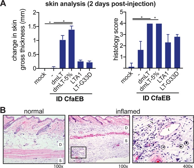
FIG 6.
Acute skin reactogenicity is altered by LT antigen after intradermal immunization. Mice (n = 3 per group) were immunized with PBS buffer alone (mock) or with PBS buffer containing 10 μg CfaEB with or without dmLT, dmLT in 5% lactose buffer, LTA1, or LT-G33D by i.d. delivery (all at 0.1 μg, except for 1 μg for LTA1). Skin samples were collected at 2 days postimmunization. (A) Changes to skin gross thickness, measured by the use of digital calipers, between the injection site and the adjacent back skin. The histology score for the severity of inflammation or tissue lesion is for H&E-stained skin sections and ranges from 0 to 5. Significance compares all groups to the naive or CfaEB i.d. groups and was determined using one-way ANOVA with the Kruskal-Wallis test with Dunn’s posttest. *, P ≤ 0.05. (B) Representative normal and inflamed injection sites are shown at magnifications of ×100 and ×400. D, dermis; S, subcutis; L, dilated lymphatics; arrows, neutrophil infiltrate; circles, mast cells. There were no differences in mast cells noted between groups.

