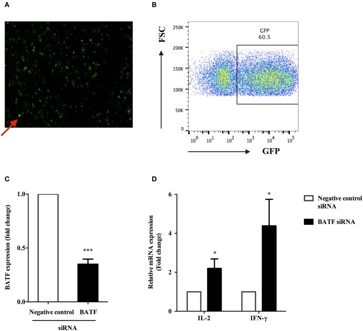
Figure 4.
BATF silencing enhanced cytokine secretions by PPD-specific T cells in PBMCs in patients with ATB. (A) Representative fluorescence microscopy image of transfected primary human PBMCs after 24 h of electroporation with 2 μg GFP Vector, and red arrow indicated transfected PBMCs containing GFP (× 200). (B) Transfection efficiency of primary human PBMCs 24 h post electroporation were assessed by analysis of GFP protein expression by flow cytometry. (C) Silencing of BATF by a BATF siRNA SMART pool in primary human PBMCs from ATB patients measured by real-time quantitative PCR. Expression normalized to a housekeeping gene (GAPDH) is presented as fold change relative to negative control siRNA. (D) PBMCs were electroporated with indicated siRNA, then cultured with M. tb-specific antigen PPD (25 μg/mL) overnight, and IL-2 or IFN-γ mRNA levels were measured by real-time quantitative PCR. The information of controls and cell viability after transfection was provided in the Methods section BATF Small Interfering RNA (siRNA) Knockdown in ATB Patients (11). Data are expressed as mean ± SEM (n = 7). *P < 0.05, ***P < 0.001. PPD, purified protein derivative; PBMCs, peripheral blood mononuclear cells; ATB, active tuberculosis; siRNA, small interfering RNA.

