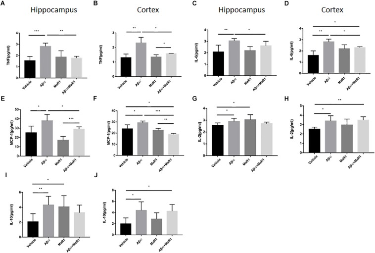
FIGURE 4.
Effects of MaR1 on the secretion of inflammatory cytokines. ELISA and CBA analysis for different inflammatory mediators in the hippocampus and cortex, TNF (A,B), IL-6 (C,D), MCP-1 (E,F), IL-2 (G,H), IL-10 (I,J). (A–F) The levels of TNF-α, IL-6 and MCP-1 in Aβ42 group were significantly higher than those in Vehicle group (P < 0.05), while treatment of Aβ42 + MaR1 significantly decreased the levels of TNF-α, IL-6 and MCP-1 induced by Aβ42 (P < 0.05). (G–J) The levels of IL-2 and IL-10 in Aβ42 group were significantly higher than those in Vehicle group (P < 0.05); MaR1 treatment increased the production of IL-2 and IL-10 in the hippocampus (P < 0.05); the levels of IL-2 and IL-10 in Aβ42 + MaR1 group were higher than those in Vehicle group in the cortex (P < 0.05). Data are expressed as mean ± SEM, 10 mice in each group, and statistical significance is defined as follows: ∗P < 0.05, ∗∗P < 0.01, ∗∗∗P < 0.001.

