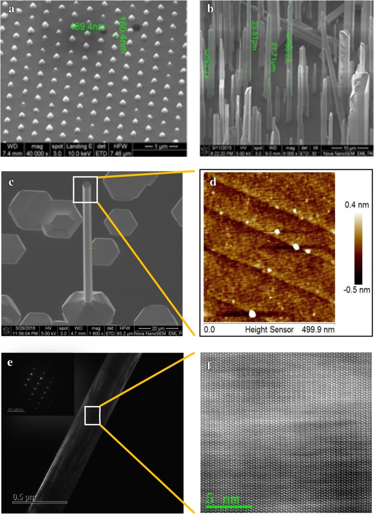
Fig. 1.
Typical scanning electron microscope (SEM) images of GaN nanocolumns grown by a selection growth method and b self-organized method, c SEM image of the self-organized catalytic-free growth GaN nanocolumn, d AFM image of the top of the GaN nanocolumn, e transmission electron microscope (TEM) image and electron diffraction pattern, f high resolution TEM atomic image

