Fig. 6.
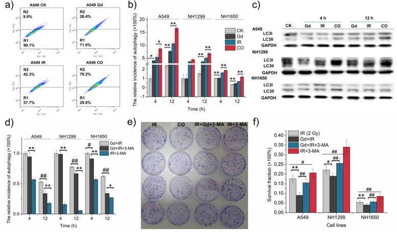
Autophagy-induced death was promoted by GONs in NSCLC cells exposed to carbon ion irradiation. a Images of autophagic rates in A549 cells at 12 h post-radiation as measured by flow cytometry (Sysmex CyFlow Cube 6, German). b The relative incidence of autophagy in the three studied cell lines. c Western blot results of LC3II expression in the three studied cell lines. d The relative incidence of GONs-induced autophagy in the presence or absence of 3-MA. e The surviving A549 cells were dyed with crystal violet in the clonogenic survival assay. f Surviving fraction of NSCLC cells after treatment with 2 Gy carbon ion radiation and/ or GONs with or without 3-MA. *p < 0.05 or **p < 0.01 represent statistically significant or extremely significant differences, respectively, induced by GONs. Similarly, #p < 0.05 or ##p < 0.01 indicated the differences owing to 3-MA treatment

