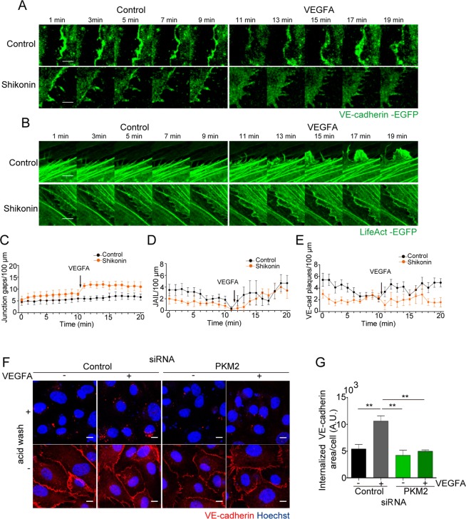
Figure 7.
PKM2 is required for junction dynamics and VE-cadherin internalization. (A and B) Still images from live microscopy of HUVECs transduced with lentivirus coding for VE-cadherin-EGFP (A) or LifeAct-EGFP (B) for 10 min prior and after addition of 20 ng/ml of VEGFA; untreated and shikonin-treated cells are shown. Scale bar, 5 µm. (C) Number of inter-cellular gaps at junctions of VE-cadherin-transduced HUVEC along time. (D and E) Number of JAIL and of VE-cadherin plaques at the lateral junctions of LifeAct-EGFP and VE-cadherin-EGFP-transduced HUVECs, respectively; means ± SEM, n = 6 junctions for 2 independent experiments. (F) Immunofluorescence of VE-cadherin (red) and Hoechst (blue, nuclei) before or after acid wash in HUVECs transfected with control or PKM2 siRNA and treated with 20 ng/ml VEGFA for 15 minutes. Scale bar, 10 µm. (G) Internalized VE-cadherin positive area per number of cells in arbitrary units (A.U.); n = 3 independent experiments, **p < 0.01 by one-way ANOVA with Sidak post-test. See related Figure S6 and Movies S5–S8 and S11–S14.

