Figure 3.
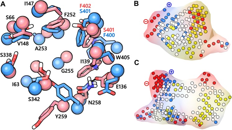
(A) The binding site of LAT1 in the outward-occluded conformation (red) superposed to the binding site of LAT1 in the inward-open state (PDB ID: 6IRT) (blue). The Cα atoms and the side chains are shown in space-filling and stick style, respectively. N258 and E136 in the outward-occluded model are engaged in a hydrogen bond interaction (black dotted line), possibly indicating a closed distal gate. While the side chain of E136 is oriented away from the binding site in the inward-open structure, suggesting an open distal gate permitting substrate access to the binding site from the cytoplasm. (B,C) are the negative images of the substrate binding cavity of the outward-occluded and the inward-open structure of LAT1, where the red, blue, yellow and white spheres are the site points corresponding to the hydrogen bond acceptor, hydrogen bond donor, hydrophobic and mixed character regions, respectively. The positive and the negative pole of the binding site are marked.

