Fig. 3.
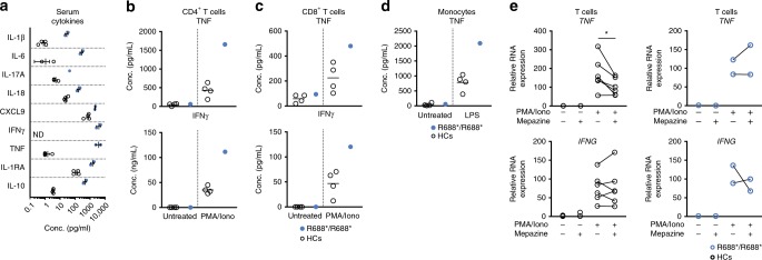
T cells and monocytes contribute to hypercytokinemia in the R688*/R688* proband. a Serum concentration of the cytokines IL-1β, IL-1RA, IL-6, IL-10, IL-17A, IL-18, IFNγ, CXCL9, and TNF in HCs (n = 4) and proband (two biological replicates) or HCs (n = 3) and proband (one biological replicate) in the case of the cytokine IL-17A. Mean and SEM are depicted. b, c ELISA of TNF and IFNγ produced by in vitro PMA/ionomycin stimulated CD4+ T cells (b) or CD8+ T cells (c) of HCs (n = 4) and proband. d ELISA of TNF produced by monocytes of HCs (N = 4) or proband treated in vitro overnight with LPS. e RT-qPCR quantifying TNF and IFNG transcripts in PMA/ionomycin stimulated T cells in absence or presence of mepazine pretreatment (20′). Cells were sampled 1 h after stimulation. Data was normalized using the housekeeping genes HPRT and GAPDH. HCs (n = 6). *p < 0.05 (paired t-test). R688* proband (n = 2). Data shown are accumulated from two independent experiments (a, e) or representative for two independent experiments (b–d). Source data are provided as a Source Data file

