Fig. 6.
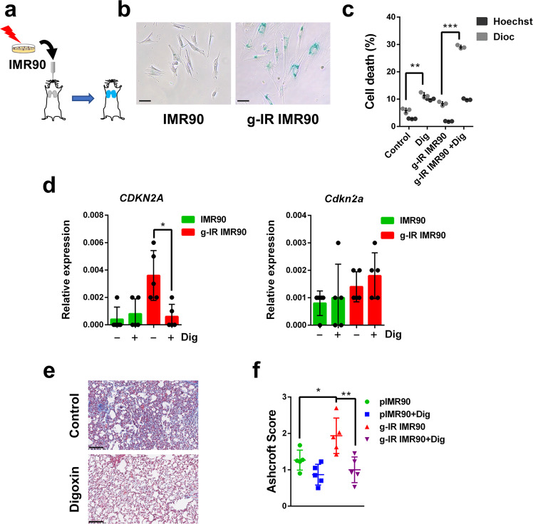
Senolytic activity of Digoxin in lung fibrosis. a Schematic diagram of the experimental system to induce lung fibrosis in mice by intratracheal instillation of proliferative or senescent gamma-irradiated IMR90 cells. b In vitro SABG staining of control or gamma-irradiated (g-IR) IMR90 cells (scale bar = 100 μm). c In vitro analysis of cell death in control or (g-IR) IMR90 cells treated or not with Digoxin (Dig). Black bars represent the % of DiOC6(3) low and grey bars represent Hoechst 33342 positive cells. n = 3 independent experiments. d Relative expression of the mRNA coding for CDKN2A (left panel) or Cdkn2a (right panel) in lung cell extracts from mice injected with control proliferative (green) or gamma-irradiated (g-IR, red) IMR90 cells, and treated or not with Digoxin, as indicated. n = 5 independent experiments. e Representative images of lung sections stained with Masson Trichrome from mice injected with gamma-irradiated IMR90 cells and treated (bottom panel) or not (upper panel) with Digoxin (scale bar = 100 μm). f Ashcroft score of Masson Trichrome staining in sections from mice injected with control proliferative or gamma-irradiated (g-IR) IMR90 cells, treated or not with Digoxin (+Dig). n = 5 independent experiments. Statistical significance was assessed by the two-tailed Student's t-test: ***p < 0.001; **p < 0.01; *p < 0.05. Data correspond to the average ± s.d. Source data for these experiments are provided as a Source Data file

