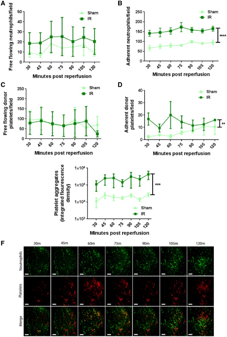
Figure 1.
Neutrophil recruitment and microthrombus formation increases rapidly following myocardial IRI. (A) Free flowing neutrophil number does not increase following IRI when compared with sham controls (n = 5/group). (B) Neutrophil adhesion within sham hearts is high but increases following IRI (****P < 0.0001 IR vs. Sham with significant differences at all time points; n = 5/group). (C) Free flowing donor platelets transmitting through myocardium is not increased following IRI (n = 5/group). (D) Singular donor platelet adhesion increases across the imaging period following IRI (**P < 0.01 IR vs. Sham; n = 5/group). (E) Accumulation of endogenous platelets, appearing as platelet aggregates and microthrombi increases following IRI and is quantitated as integrated fluorescence density (***P < 0.001 IR vs. Sham with significant differences at all time points; n = 5/group). Two-way ANOVA with Sidak’s multiple comparison test used for all analysis. (F) Representative intravital images of the beating mouse heart to show rapid accumulation and gradual increases of both neutrophils and platelets. Neutrophils present primarily as individual cells with platelets found as aggregates or microthrombi. Merged images show aggregates comprised of both cell types (yellow). Green, Neutrophils (PE+anti-Gr-1ab); Red, endogenous platelets (APC+anti-CD41ab). Scale bars represent 100 µm.

