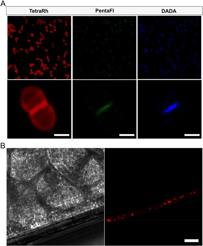
Figure 5.

(A) Confocal microscopy image of E. faecium (WT) treated with 5 min pulse of 500 μM TetraRh, 500 μM PentaFl, and 5 mM DADA (scale bar: 1 μm). (B) In vivo labeling of E. faecium in model host. C. elegans were infected with E. faecium for 4 h, washed to remove noncolonized bacteria, and incubated with 50 μM TetraRh for 2 h. The C. elegans were washed, anesthetized, mounted on a bed of agarose, and imaged using confocal microscopy (scale bar: 10 μm).
