Figure 3.

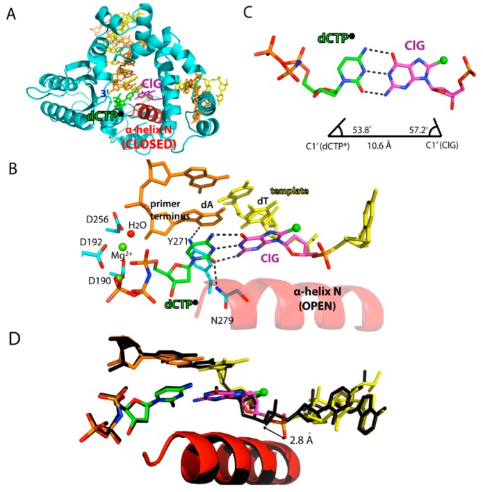
Ternary structure of polβ incorporating non-hydrolyzable dCTP analog opposite templating ClG. (A) Overall view of the polβ-ClG:dCTP* ternary complex structure. ClG is shown in magenta and the incoming dCTP* is colored green. The α-helix N, which is shown in red, forms a closed conformation. (B) Active-site view of the polβ-ClG:dCTP* ternary complex structure. The templating ClG is an anti-conformation and forms three Watson-Crick hydrogen bonds with incoming dCTP*. The minor groove edges of the incoming dCTP and primer terminus are recognized by Asn279 and Tyr271, respectively. Hydrogen bonds are indicated in dashed lines. Metal cofactors are shown in green spheres and water is shown in red sphere. (C) Base pair geometry of dCTP*:ClG in the active site of polβ. (D) Superposition of the polβ-ClG:dCTP* structure (multicolor) with the polβ-dA:dUTP* ternary complex structure (black). The distance between the 5′-phosphate oxygen of the templating bases is shown.
