Figure 5.
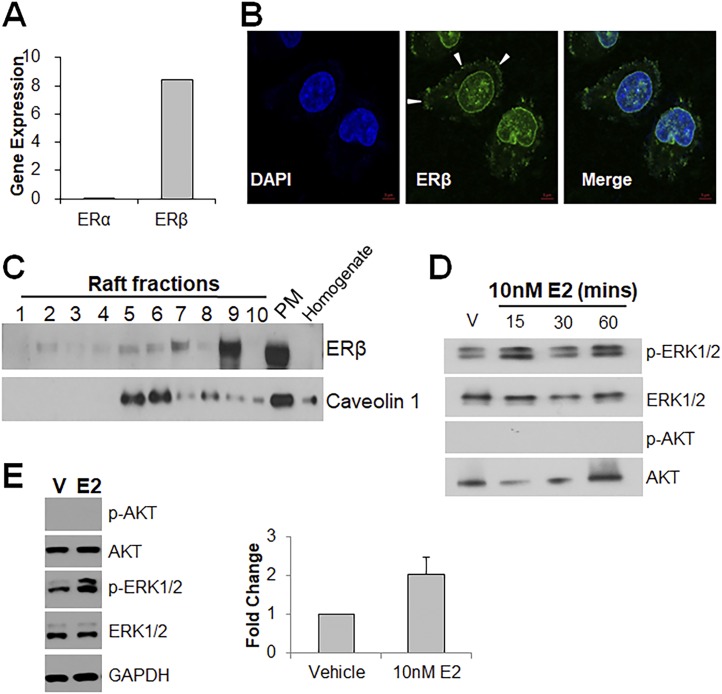
Prostate cancer stem-like cells express ERβ on their membranes and signal through the MAPK pathway. (A) qRT-PCR shows high expression of ERβ mRNA and an absence of ERα mRNA in HuSLCs. (B) Immunocytochemistry of HuSLCs for ERβ shows intracellular, nuclear, and membrane-bound (arrowheads) ERβ protein (green). Nuclei were stained with DAPI (blue). Magnification ×40. (C) Membrane fractions of HuSLCs were separated by density gradient centrifugation followed by western blot analysis for ERβ protein. ERβ was found at high levels in the whole plasma membrane fraction (PM) and the lipid raft fractions (5–8) marked by caveolin-1 expression. (D) HuSLCs treated with 10 nM E2 for 15, 30, and 60 min showed AKT phosphorylation but no activation of ERK1/2. (E) Prostate cancer stem/progenitor cells were isolated from DU145 cultures by transferring to three-dimensional Matrigel culture for 3 wk. Harvested DU145 spheres were exposed to 10 nM E2 for 30 min. Western blot analysis showed an increased ERK1/2 phosphorylation but no AKT activation as compared with vehicle-treated controls. Graphic representation of p-ERK1/2 signal at right for n =3.

