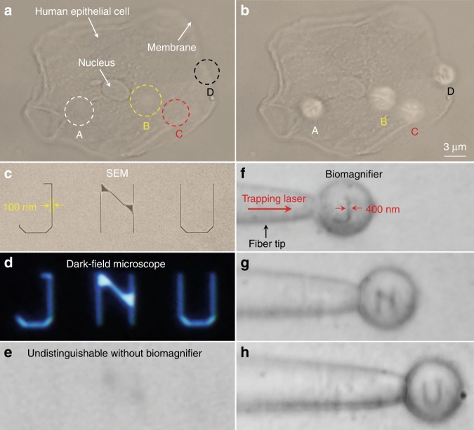
Fig. 3. Nano-optical imaging of subcellular structures and nanopatterned letters.
a, b Optical images of the subcellular structures of a human epithelial cell using a conventional optical microscope (a) and biomagnifiers (b). The positions of four biomagnifiers are marked as A–D. For comparison, the biomagnifiers can resolve the fibrous cytoskeleton (indicated as A–C) inside the cell and two-layer structures (indicated as D) on the cell membrane, which are indistinguishable by the conventional microscope. c–e SEM (c), dark-field (d), and optical images (e) of nanopatterned letters JNU, which represent the acronym of Jinan University. The line width of the nanopatterned letters is 100 nm, which is smaller than the diffraction-limit resolution of the conventional optical microscope. f–h Optical images showing that the biomagnifier trapped on the fiber tip can scan and image the nanopatterned letters J (f), N (g), and U (h) by moving the fiber. The line width of the nanopatterned letters was magnified from 100 to 400 nm

