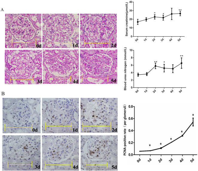
Fig. 1.
Periodic acid-Schiff (PAS) staining and proliferating cell nuclear antigen (PCNA) immunohistochemical staining during the development of anti-Thy1 nephritis. a PAS staining, and serum creatinine and blood nitrogen levels. b PCNA immunohistochemical staining. The percentage of PCNA-positive cells was calculated as the number of positive cells relative to the number of total glomerular cells (10–15 glomeruli were counted for each rat). Scale bars=10 μm. 0 d: Control group: 1–5 d: Days after the injection of the anti-Thy1 antibody. *p < 0.05, **p < 0.01; n = 4

