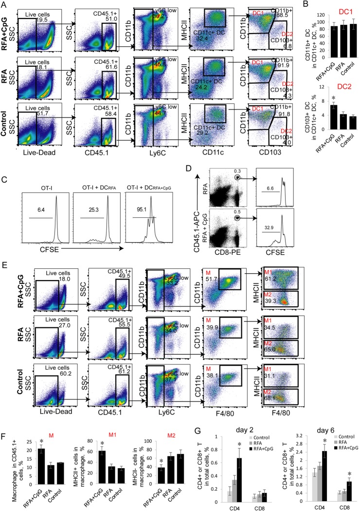
Fig. 4.
Administration of adjuvant CpG increases the frequency of tumor-associated immunogenic DC2 and M1 and enhances T-cell tumor infiltration. a, b Flow cytometry analysis of tumor-associated DCs in tumor single cell suspensions derived from EG7 tumors in B6.1 mice was performed with a progressive gating strategy. The frequency of tumor-associated CD11b+CD11c+ DC1 and CD11c+CD103+ DC2 in the total CD11c+ DC population at 2 days post treatment is shown; n = 3 mice per group. *P < 0.05 versus the cohort of control mice (Student’s t-test). c In vitro T-cell proliferation assay. Irradiated (2000 rad) DCs that had been initially purified from the tumor-draining lymph node cells of RFA– and RFA+ CpG-treated tumor-bearing mice were cultured with CFSE-labeled OT-I CD8+ T cells for 3 days and analyzed for T-cell proliferation by flow cytometry. The value represents the percentage of cells that divided at least once. d In vivo T-cell proliferation assay. Cells from the tail blood samples of RFA- and RFA/CpG-treated EG7 tumor-bearing mice (4 each group) with transferred CFSE-labeled OT-I CD8+ T cells were stained with anti-CD45.1-APC and anti-CD8-PE antibodies and analyzed by flow cytometry. CD45.1/CD8 double-positive T cells were gated for the analysis of T-cell proliferation. The value represents the percentage of cells that divided more than three times. e, f Flow cytometry analysis of tumor-associated macrophages from the tumor single cell suspensions derived from EG7 tumors in B6.1 mice by a progressive gating strategy. The frequency of tumor-associated CD11b+F4/80+ macrophages (M) in the total CD45.1+ cell population as well as the frequency of tumor-associated inflammatory CD11b+F4/80+Iab+ M1 and tolerogenic CD11b+F4/80+Iab- M2 in the total CD11b+F4/80+ M population at day 2 post treatment; n = 3 mice per group. *P < 0.05 versus the cohort of control mice (Student’s t-test). g The frequency of CD4+ and CD8+ T cells in the total cell population at day 2 and day 6 post treatment; n = 5 mice per group. Tumor-infiltrating T cells were gated from CD45.1+CD3+ cells and analyzed for the expression of CD4 and CD8 by flow cytometry. *P < 0.05 versus the cohort of control RFA mice (Student’s t-test). One representative experiment out of two experiments is shown

