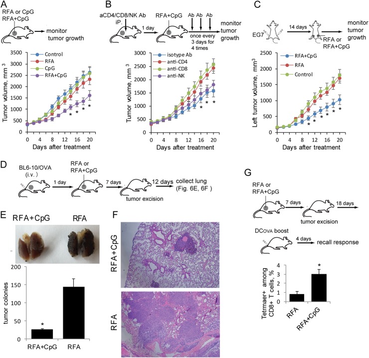
Fig. 6.
Combination treatment with RFA and CpG leads to the inhibition of larger tumor growth and lung tumor metastasis. a A schematic diagram of the RFA, CpG and RFA+CpG treatments for mice bearing larger EG7 tumors. Mice bearing larger EG7 tumors(8 per group) were treated with RFA, CpG, RFA+CpG or the control PBS. Tumor growth or regression was monitored. *P < 0.05 versus the cohort of the control or CpG-treated mice (Student’s t-test). b Experimental setup of the RFA+CpG treatment of mice bearing larger EG7 tumors with or without the depletion of CD4+ T, CD8+ T and NK cells. To assess the involvement of CD4+ T, CD8+ T and NK cells in the RFA+CpG-induced inhibition of tumor growth, mice bearing larger EG7 tumors (8 per group) were injected with anti-CD4, anti-CD8 or anti-NK Abs for the depletion of CD4+ or CD8+ T cells or NK cells, respectively. Mice were then treated with RFA+CpG therapy one day after the antibody injection. The antibody injection was repeated once every 3 days for a total of 4 injections. Tumor growth or regression was monitored. *P < 0.05 versus the cohort of CD4+ or CD8+ T cell-depleted mice (Student’s t-test). c C57BL/6 mice were injected subcutaneously with EG7 cells (2 × 10⁶ and 4 × 10⁶) on the left and right sides of the lower back, respectively. When the right and left tumors reached ~350 mm3 and ~200 mm3, respectively, RFA+CpG or RFA treatment was performed on only the right tumor. The growth of the left and right site tumors was monitored. For ethical reasons, mice were killed when the right site tumor reached ~2500 mm3. The sizes of the left untreated tumors in the different groups were compared. *P < 0.05 versus the cohort of untreated control mice. d A schematic diagram of the experiments assessing the antimetastatic activity of the RFA+CpG treatment in mice bearing larger tumors. Mice with larger s.c. EG7 tumors (8 per group) were i.v. injected with BL6–10OVA cells. One day later, RFA or RFA+CpG was performed. The s.c. tumors were surgically removed 7 days post RFA therapy. The mice were killed 20 days post BL6–10OVA cell injection, and the lungs were collected. e Metastatic black BL6–10OVA tumor colonies in the lungs of the mice (5 per group) were counted. *P < 0.05 versus the cohort of RFA-treated mice (Student’s t-test). f The lungs were sectioned and stained with hematoxylin and eosin. g A schematic diagram of the experiments assessing the memory T cells produced by RFA+CpG treatment in mice bearing large tumors. Mice bearing larger s.c. EG7 tumors (6 per group) were treated with RFA or RFA+CpG. The tumors were surgically removed 7 days post RFA therapy. Mice were boosted with DCOVA 25 days post RFA therapy, and the memory T-cell recall responses were assessed 4 days after the boost by flow cytometry. *P < 0.05 versus the cohort of RFA-treated mice (Student’s t-test). One representative experiment out of two experiments is shown

