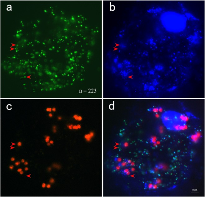
Fig. 4. Stained protoplasts isolated from melon.
Leaves of melon transformed with a mitochondrially targeted GFP (35S-coxIV-GFP) were stained with DAPI and visualized at excitation wavelengths for green fluorescent protein (a) and DAPI (b). The number of mitochondria was 223, as determined by ImageJ software. Red autofluorescence was observed from the chloroplasts (c). It is clear that mtDNA signals were not present in a portion of the mitochondria after image merging (d). The red arrows highlight mitochondria without mtDNA signals. Bar = 10 μm

