Figure 3.
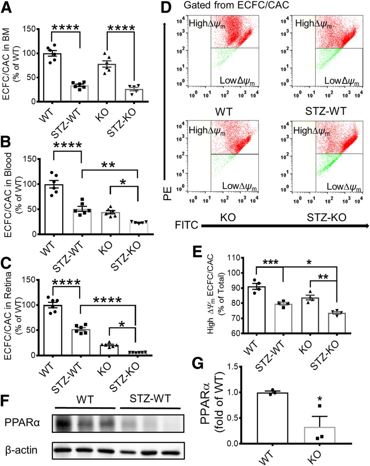
PPARα KO decreased ECFC/CAC numbers, homing, and mitochondrial function in diabetic mice. ECFC/CAC numbers were quantified in the BM (A), blood (B), and retina (C) of STZ-induced diabetic WT mice (STZ-WT) at 16 weeks after the onset of diabetes, their age- and genetic background–matched nondiabetic WT mice (WT), STZ-induced diabetic PPARα−/− mice (STZ-KO), and nondiabetic PPARα−/− mice (KO) (n = 5). D and E: ∆Ψm of ECFC/CACs in the blood was measured by FCM, and the percentage of high ∆Ψm was compared between the groups as indicated. D: Representative FCM profiles. E: The percentage of high ∆Ψm cells was quantified and compared among the four groups (n = 4). Representative Western blots (F) and densitometry analyses (G) of PPARα protein levels in the BM of WT mice and STZ-induced WT mice (mean ± SEM; *P < 0.05; **P < 0.01; ***P < 0.001; ****P < 0.0001). PE, phycoerythrin.

