Fig. 3.
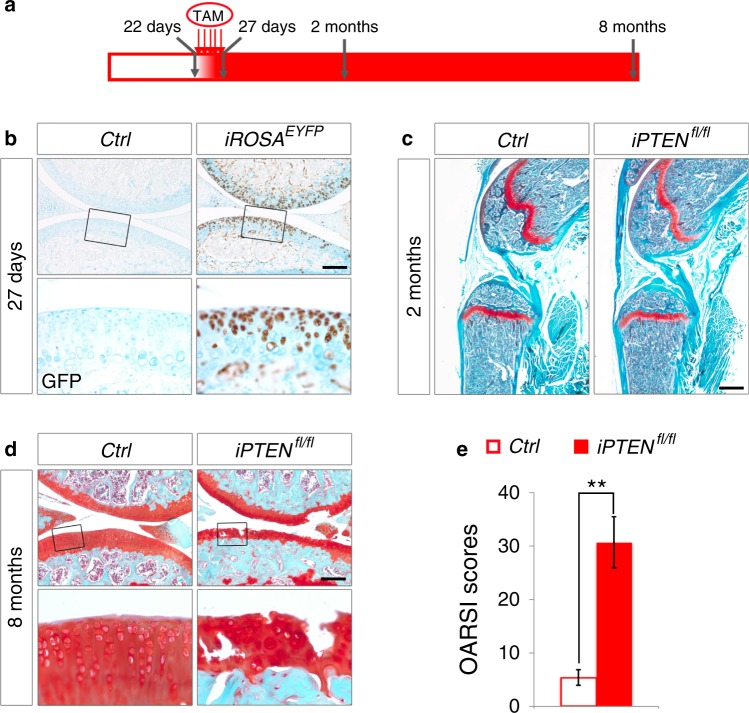
Induced PTEN deficiency in adult articular chondrocytes causes OA phenotypes in mice. a Schematic diagram showing the protocol of tamoxifen administration for starting ROSAEYFP expression or ablating the PTEN gene in articular chondrocytes. Five successive doses of tamoxifen were injected every day since 22-day-old. Knee joints were analyzed at 27 days, 2 months and 8 months of age. b Representative images of immunohistochemical staining of EYFP in articular cartilage from 27-day-old Ctrl and iROSAEYFP mice (n = 3 per group). Tamoxifen was injected since 22-day-old. The framed area in each picture is shown below at a higher magnification. c Representative images of safranin O staining of hind limbs from 2-month-old Ctrl and iPTENfl/fl mice (n = 4 per group). Tamoxifen was injected since 22-day-old. d Representative images of safranin O staining of knee joints from 8-month-old Ctrl and iPTENfl/fl mice (n = 12 per group). Tamoxifen was injected since 22-day-old. The framed area in each picture is shown below at a higher magnification. e Quantified pathological changes of each group at 8 months. Each value represents the mean ± SEM (n = 12 per group). **P < 0.01. Scale bars: 250 µm in (b) and (d), 800 µm in (c)

