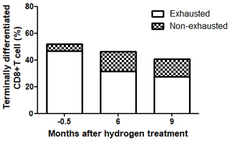
Figure 4.
In terminally differentiated CD8+ T cells, the proportion of exhausted cells varied with the treatment time. The results were tested by flow cytometry, in which the terminally differentiated CD8+ T cells were labeled CD3+CD8+CD27-, in which PD-1+, was considered a marker of exhaustion and non-exhausted cells were PD-1.

