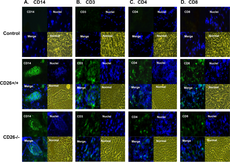
Fig. 7. Determination of infiltrated macrophages and T lymphocytes in grafts of CD26+/+ and CD26–/– mice after skin transplantation.
The tail skin before transplantation was collected as a control; graft tissues of CD26+/+ and CD26–/– mice were obtained on day 7 after transplantation. The frozen sections of skin graft were stained with monoclonal antibody against mouse CD14, CD3, CD4, or CD8, and the nucleus was then counterstained with Hoechst 33,342. a–d Immunofluorescence analysis results of infiltrated CD14+ cells, CD3+ cells, CD4+ cells, or CD8+ cells in the grafts of CD26+/+ and CD26–/– mice, respectively. Photographs are shown at 400× magnification

