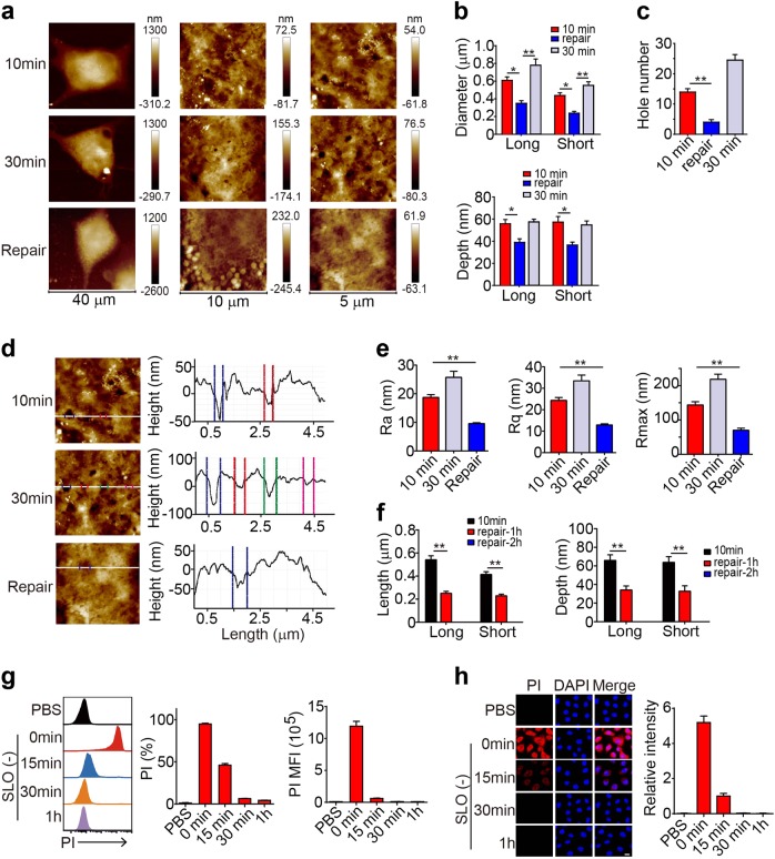
Fig. 3.
SLO-induced pore formation is repairable. a–e OVA-B16 cells were treated with 50 U of SLO for 10 min or 30 min. Some cells were treated with SLO for 10 min and then cultured in the absence of SLO for another 20 min. Cells were fixed and imaged using AFM. Representative images of OVA-B16 cell topographies at different magnifications are shown (a). The pore diameter and depth were measured (b). The number of pores was counted within three 10 × 10 μm2 areas from one cell (n = 6) (c). Surface roughness was analyzed (d), and the values of Ra, Rq, and Rmax were calculated (e). f OVA-B16 cells were treated with SLO (50 U) for 10 min, and then cultured in fresh medium without SLO for an additional 1 h or 2 h. Cells were imaged using AFM to determine the pore diameter and depth. (g and h) OVA-B16 cells were treated with SLO (50 U) for 10 min, and then cultured in SLO-free medium for another 15 min, 30 min, or 1 h, as indicated. During the last 30 s of repair, PI was added to the culture medium. PI+ cells were measured by flow cytometry (g) and confocal microscopy (h). Bar, 20 μm. * p < 0.05 and ** p < 0.01 by one-way ANOVA (b, c, e and f). The data are presented as the means ± SEM of three independent experiments

