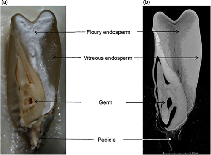
Figure 6.

Comparison of a longitudinal digital image and CT image slice. (a) A longitudinal digital image; (b) 2D CT image slice (Guelpa et al., 2015). The voxel size, voltage, and scan time are 13.4 μm, 60 kV, and 30 min, respectively. The same maize grain is depicting the internal structure of the maize grain, that is, flour and vitreous endosperm, germ, and pedicle. In CT image, the brighter gray region represents the denser vitreous endosperm and the darker region the less dense floury endosperm. The vitreous endosperm thus appeared translucent (Figure a) due to no light being reflected
