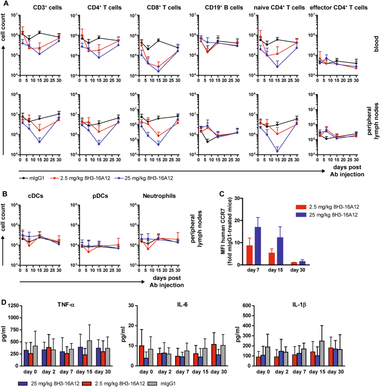
Fig. 4.
Single dose toxicity study of anti-human CCR7 mAb 8H3-16A12. GL-CCR7+/+ mice received a single i.p. injection of 2.5 mg/kg 8H3-16A12, 25 mg/kg 8H3-16A12 or 25 mg/kg mIgG1 control Ab and were sacrificed 2, 7, 15, and 30 days post-injection. The cell counts of the indicated immune cell subsets (a, b) were assessed in the blood and pLN by flow cytometry. The data are presented as the mean ± SD from 5–8 mice per group. c Coating of human CCR7 by 8H3-16A12 on T cells in blood. Cells were stained with anti-mIgG1 Ab, and the MFI of the staining is presented as fold change over mIgG1-treated mice. The data are presented as the mean ± SD from 3–7 mice per group. d Levels of TNF-α, IL-6, and IL-1β were measured in sera in a Luminex-based assay. The data are presented as the mean ± SD from 6–9 per group

