Fig. 1.
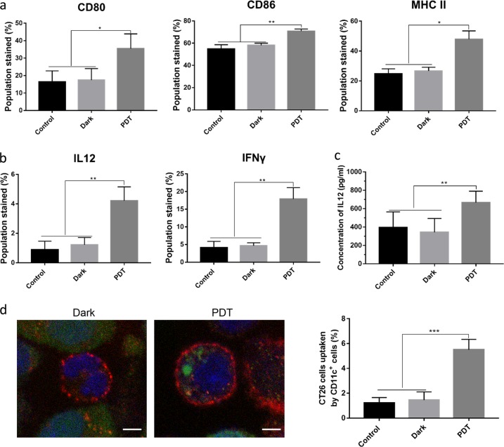
Activation of dendritic cells. Dendritic cells were cocultured with BAM-SiPc-PDT-treated CT26 cells for 24 h before the following analyses were performed. a Cells were collected and stained with anti-CD11c FITC-conjugated, anti-CD80 PE-conjugated, anti-CD86 APC-conjugated, and anti-MHC II PE-conjugated antibodies. The percentage of CD11c-positive dendritic cells with high expression of CD80, CD86, and MHC II was calculated after flow cytometric analysis (N = 5). b Five hours after the addition of Brefeldin A (10 μM), cells were stained with anti-CD11c FITC-conjugated antibodies together with either anti-IL12 APC-conjugated or anti-IFNγ APC-conjugated antibodies before flow cytometry was carried out to examine the expression of these cytokines (N = 4). c Supernatants were collected after coculture to determine the concentration of IL12 by ELISA (N = 3). d The engulfment of CT26 cells by dendritic cells was assessed. PDT-treated CT26 cells were stained with CMFDA (green) and then incubated with dendritic cells for 24 h. The cells were collected and stained with anti-CD11c antibodies (red). The left panel shows the engulfment of the CT26 cells (green) by the dendritic cells (red), which was visualized by confocal microscopy. The nucleus was stained in blue, bar = 5 μm (N = 3). The right panel shows the percentage of CT26 cells engulfed, which was estimated by flow cytometry (N = 5). *p < 0.05; **p < 0.005; ***p < 0.001

