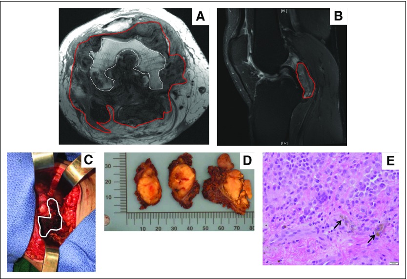
Fig 3.
Pigmented villonodular synovitis.(A) Axial T1-weighted magnetic resonanceI image that shows lobulated PVNS (red line) throughout joint and extensive subjacent erosions of femoral condyles (white outline). (B) Sagittal T2 fat-saturated magnetic resonance image that shows a multilobulated intra-articular mass abutting the posterior cruciate ligament, tibial plateau, and posterior knee joint capsule. (C) Intraoperative imaging that shows a brownish pigmented mass. (D) Gossly resected specimens that show multiple yellowish to brown masses with no encapsulation. (E) Randomly distributed multinucleated osteoclast-like giant cells accompanied by mononuclear cells, histiocytes, and hemosiderin-laden macrophages (black arrows; magnification, 200×). Images courtesy of David Panicek, MD, Department of Radiology, Meera Hameed, MD, Department of Pathology, and Nicola Fabbri, MD, Department of Orthopedic Oncology Surgery, Memorial Sloan Kettering Cancer Center, New York, NY.

