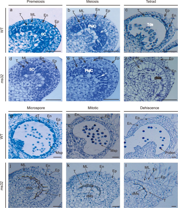
Fig. 2. Histological examination of anthers at different developmental stages in WT and ms32 plants.
Transverse sections of WT (a–c, g–i) and ms32 (d–f, j–l) anthers at different developmental stages. a, d Premeiotic stage; b, e Meiotic stage, with white arrows pointing to the crushed and separated PMCs; c, f Tetrad stage; g, j Microspore stage; h, k Mitotic stage; i, l Dehiscence stage. dMs, degenerated meiocytes; dT, degenerated tapetum; En, endothecium; Ep, epidermis; ML, middle cell layer; Msp, microspore; PMC, pollen mother cell; SC, sporogenous cell; T, tapetum; Tds, tetrads. Scale bars, 50 μm

