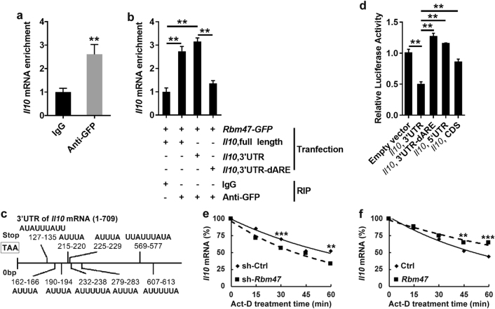
Fig. 3.
Interaction between Rbm47 and Il10 mRNA. a, b HEK-293T cells were transfected with GFP- or Rbm47-GFP-expressing plasmid (a). HEK-293T cells were transfected with plasmids containing Rbm47-GFP with Il10, full-length, Il10, 3′UTR or Il10, 3′UTR-dARE (b). After 2 days, the cells were harvested and subjected to RIP with IgG and anti-GFP antibody. The precipitated mRNA was then reverse transcribed into cDNA and subjected to q-PCR analysis. c AREs in the 3′UTR of Il10 mRNA. d HEK-293T cells were co-transfected with pGL3 containing Il10 promoter and Rbm47-expressing or empty vector. After 48 h, the cells were subjected to the dual-luciferase reporter assay. Relative luciferase activity was counted (first fluorescence intensity/second fluorescence intensity). e SP2/0 cells were infected with sh-Rbm47-expressing or control lentivirus and treated with actinomycin D (10 mg/ml). Total RNA was harvested at 0, 15, 30, 45, or 60 min after treatment. f Il10-expressing plasmids were co-transfected with Rbm47-expressing or empty plasmids into CHO cells, and then the cells were treated with actinomycin D (10 mg/ml). Total RNA was harvested at 0, 15, 30, 45, or 60 min after treatment. Il10 mRNA levels were measured using q-PCR and normalized to gadph mRNA. The normalized level of Il10 mRNA at time 0 was set to 100. Data are representative of at least three independent experiments, and error bars indicate the standard deviation. **P < 0.01; ***P < 0.001

