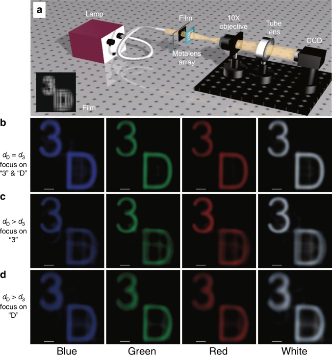
Fig. 6. Imaging demonstration of the broadband achromatic metalens array.
a Measurement configuration for the imaging demonstration of the broadband achromatic metalens array. The object films illuminated by blue, green, red and white light from the lamp and their reconstructed images can be captured by the following microscopy imaging system. Blue, green and red light is generated by inserting narrow-band (10 nm) filters of 488 nm, 532 nm and 633 nm, respectively. b Reconstructed images in the case of dD = d3, which means that the number “3” and the letter “D” are on the same depth plane relative to the central depth plane. The characters “3” and “D” will always become clear together in this case. Good-quality images, especially in the white-light case, can be observed by adjusting the film and metalens array to match each other. The white-light image explicitly shows the broadband achromatic property of the metalens array in the entire visible region. Scale bar, 100 μm. c, d Reconstructed images in the case of dD > d3, which means that the number “3” is closer to the central depth plane than the letter “D”, and they are on different depth planes (Fig. S5b shows the sketch map). Hence, when we move the microscopy imaging system along the optical axes to focus on the number “3” (as shown in c), the letter “D” continues to blur since they are not on the same depth plane. When we focus on the letter “D” in (d), the number “3” will become blurred compared with that in (c). These imaging results effectively demonstrate the three-dimensional image depth using our metalens array in integral imaging. Scale bar, 100 μm

