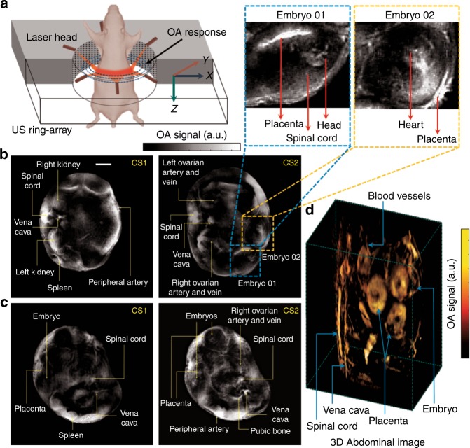
Fig. 1. In vivo whole-body imaging of pregnant mice at gestations days E14 and E19.
a Schematic of the ring-shaped optoacoustic tomography (ROAT) system. b In vivo tomographic scans of the abdominal cavity of an E19 pregnant mouse at two different cross-sections (CS1 and CS2): anatomical lay-out highlighting the different internal organs of the mother mouse along with embryos, labelled on the zoomed-in sections. c Similar cross-sections (CS1 and CS2) from an E14 pregnant mouse with anatomical phenotyping of various internal organs in the abdominal region. The scale bars for both (b, c) are 5 mm. d Three-dimensional abdominal image of an E14 mouse acquired with spiral volumetric optoacoustic tomography (SVOT)10

