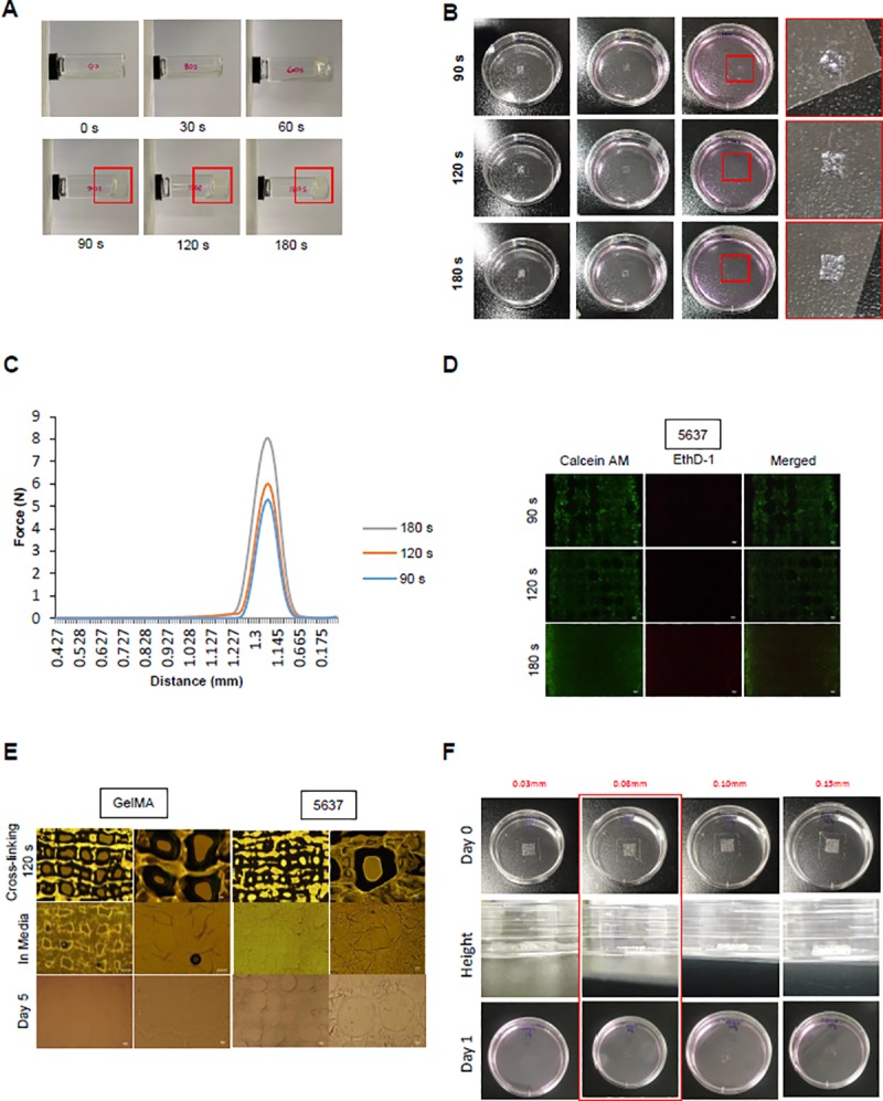
Fig 2. UV crosslinking condition for 3D culture model.

(A) After adding 1 ml to the vials, the UV crosslinking time condition was confirmed. The times were fixed at 0, 30, 60, 90, 120, and 180 s, and a 365 nm UV lamp was used. (B) The pattern was printed on a 60-mm plate using GelMA. After crosslinking at the conditions provided in (A), complete media was added and samples were incubated for 1 day in a 37°C, 5% CO2 incubator. (C) The shape of the structure was measured after 90, 120, and 180 s. The intensity force at which the structure was destroyed under the conditions of constant distance (1.225 mm) and time (0.7 s) was measured. (D) The GelMA structure and GelMA/bladder cancer cells mixture was printed and crosslinked. After being placed in complete media, the GelMA structure was confirmed with a microscope, and the structure was maintained after incubation for 5 days. (E) To investigate the effect of crosslinking time on the cells, we performed live/dead staining. After 1 day of culture, the printed mixture of GelMA and cells was treated with Calcein-AM (2 μM) and EthD-1 (4 μM) and examined by fluorescence microscopy. The shape of the structure was examined. (F) The different layers of scaffold in the Z-direction. The constructs were printed at different heights of 0.03, 0.08, 0.1, and 0.15 mm with only GelMA. Some of the structure was maintained even after 1 day in the complete media.
