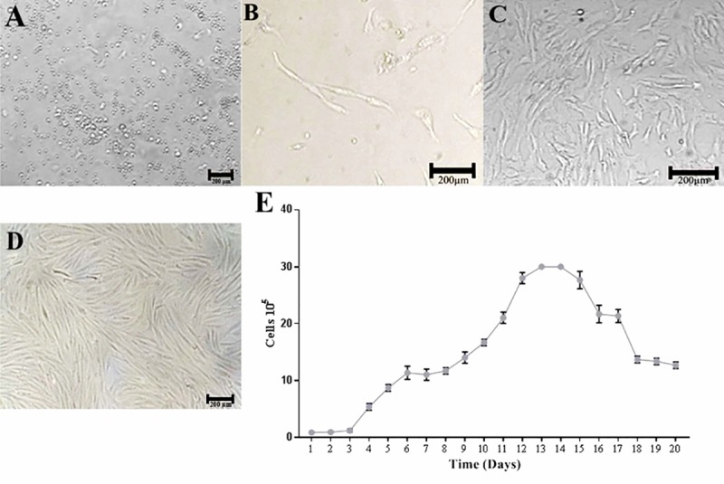
Fig 1. Photomicrography of g-ASCs cell culture isolated from subcutaneous adipose tissue of goats.

(A) Stromal fraction of adipose tissue in culture medium DMEM-F12, 24 hours of insulation, 10×10. (B) Cells with fibroblastoid plastic-adherent morphology, five days, 10×10. (C) Cells in 20 days of culture, 80% confluence, 10×10. (D) Cell culture of g-ASC, after four days of thawing, 90% confluence, 10×10. (E) Growth curve (20 days) of in vitro cultured g-ASCs.
