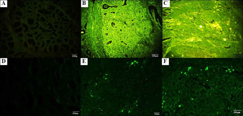
Fig 6. Photomicrography of the mammary gland of goats without staining after 30 days of therapy with g-ASCs.

(A) Parafinated tissue without marking with Qdot (control), 10 × 20. (B, C) Parafinate tissue of the right mammary gland (without g-ASCS injection), and left (with g-ASCS injection), respectively, 10x20. (D) Frozen tissue without marking with Qdot (control), 10 × 20. (E, F) Frozen tissue of the right mammary gland (without g-ASCS injection), and left (with g-ASCs injection), respectively, 10x20.
