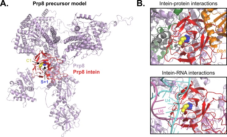
Fig 7. Modeling the Cne Prp8 intein into Prp8 and docking into the spliceosomal U4/U6.U5 tri-snRNP reveals unfavorable interactions.
(A) Cne Prp8 intein in Prp8 exteins. The Cne Prp8 intein structure (red) was modeled into a structure of S. cerevisiae (PDB 5GAN, chain A) Prp8 exteins (lavender). The S+1 is shown as blue spheres to indicate the site where a peptide bond was broken to insert the intein. The C1 is shown as yellow spheres and specifies start of the intein. The Prp8 intein localizes to a crowded site of the Prp8 structure, in a region that is highly conserved and functionally important. (B) The Cne Prp8 intein in the spliceosomal U4/U6.U5 tri-snRNP. The intein-containing Prp8 was overlaid into the tri-snRNP subunit structure (PDB 5GAN). This revealed a clash of the intein with spliceosomal protein Dib1 (top panel, orange). There are also possible clashes with the intein and U4 snRNA and U6 snRNA (bottom panel, teal and pink, respectively). Cne, C. neoformans; PDB, Protein Data Bank; Prp8, pre-mRNA processing factor 8; snRNA, small nuclear RNA; tri-snRNP, triple small nuclear ribonucleoprotein.

