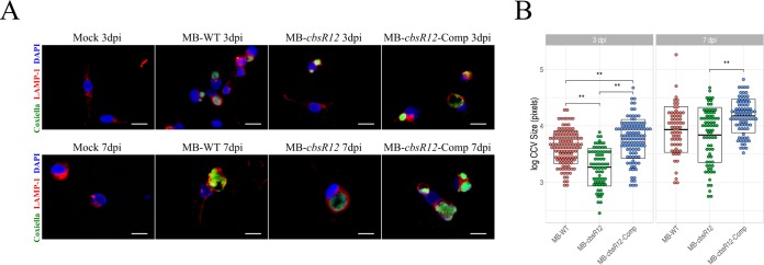
FIG 4.
CbsR12 affects CCV expansion in infected THP-1 cells. (A) Representative IFAs of MB-WT, MB-cbsR12, and MB-cbsR12-Comp infecting THP-1 cells at 3 and 7 dpi. C. burnetii was probed with anti-Com1 antibodies coupled to Alexa Fluor 488 (green), CCV boundaries were labeled with anti-LAMP1 antibodies coupled to rhodamine (red), and host cell nuclei were labeled with DAPI (blue). Scale bars, 20 μm. (B) Sizes of individual CCVs in log10 (pixels) for MB-WT, MB-cbsR12, and MB-cbsR12-Comp. Measurements were taken from 46 individual images of random fields of view spanning three different experiments for each C. burnetii strain. Crossbars represent means ± the SEM (**, P < 0.01, one-way analysis of variance).

