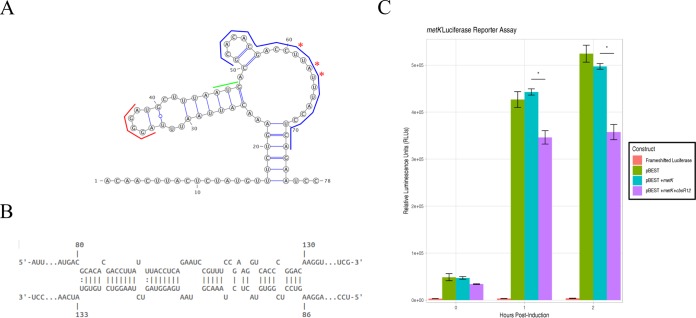
FIG 8.
CbsR12 targets and downregulates translation of a metK-luciferase fusion construct. (A) Secondary structure of the metK 5′ UTR and initial coding sequence as predicted by Mfold. Asterisks indicate apparent alternative “TSSs” determined by 5′ RACE. (Nucleotide 1 was determined to be the TSS for the full-length transcript by 5′ RACE.) Colored lines represent the start codon (green), a predicted RBS (red), and the determined CbsR12-binding site (blue). (B) Representation of CbsR12 binding to the metK transcript, as determined by IntaRNA, with the base numbers indicated. The top strand in the model represents the metK sequence, while the bottom strand represents the complementary CbsR12 sequence. (C) metK-luc reporter assay indicating relative luminescence units produced by pBESTluc constructs with (i) no luciferase production (frameshifted luciferase), (ii) pBESTluc vector (pBEST), (iii) pBESTluc with the CbsR12 binding site cloned in frame into luc but lacking the cbsR12 gene (pBEST+metK), and (iv) pBESTluc with the CbsR12 binding site cloned in frame into luc plus the cbsR12 gene driven by a Ptac promoter (pBEST+metK+cbsR12). Values represent means ± the SEM from three independent determinations (*, P < 0.05 [Student t test]).

