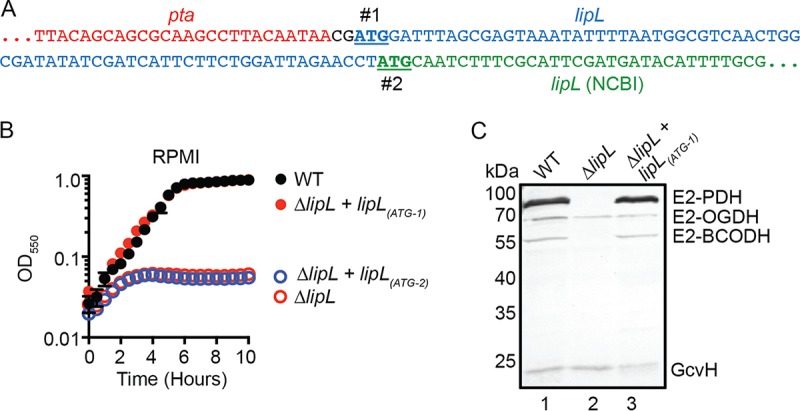
FIG 1.

The start codon of lipL is two nucleotides downstream of the pta stop codon in S. aureus. (A) DNA sequences showing two potential in-frame start codons of lipL (#1 and #2) are underlined. The 5′ end of lipL, as published by NCBI, is in green, while additional nucleotides that would form the 5′ end of lipL with the earlier start codon are in blue. (B) Growth of WT, ΔlipL mutant, ΔlipL+lipL(ATG-1) mutant, and ΔlipL+lipL(ATG-2) mutant strains in RPMI medium over 10 h. (C) In vivo lipoylation profiles of WT, ΔlipL mutant, and ΔlipL+lipL(ATG-1) mutant strains after 9 h subculture growth in RPMI medium plus BCFA and NaAc was assessed by immunoblotting SDS-PAGE-resolved whole-cell lysates with rabbit anti-lipoic acid antibody. The presented blot is representative of at least three independent experiments.
