. 2019 Oct 22;8:e45465. doi: 10.7554/eLife.45465

Figure 4. The morphology of the ciliary phaosomal cells in adult Maritigrella crozieri.

(A) A dense cluster of intra-cellular vacuoles (phaosomes – highlighted in magenta) filled with cilia (n = 3 individuals). (B) Multiple cilia in the phaosome. (C) A possible unpigmented supporting cell (asterisk) wrapping around the phaosome cell with mitochondria (m) and Golgi apparatus (ga) in the cytoplasm. (D) Ciliary axonemata (ax) are anchored in the cytoplasmic layer (cl) by basal bodies (bb). (E) 3D reconstruction of the interior of a third of a phaosome, showing that the cilia are unbranched (pink) and the basal bodies (yellow) are distributed all around the phaosome. (F) Cross sections of the ciliary axonemata show various arrangements of microtubules: 9 × 2+ two with dynein arms attached to the A-tubules (arrow), 9 + 2 singlets (double arrowheads), and singlets (triple arrowheads). This variation is related to the distance from the basal body (Figure 4—videos 3), (cm) ciliary membrane.

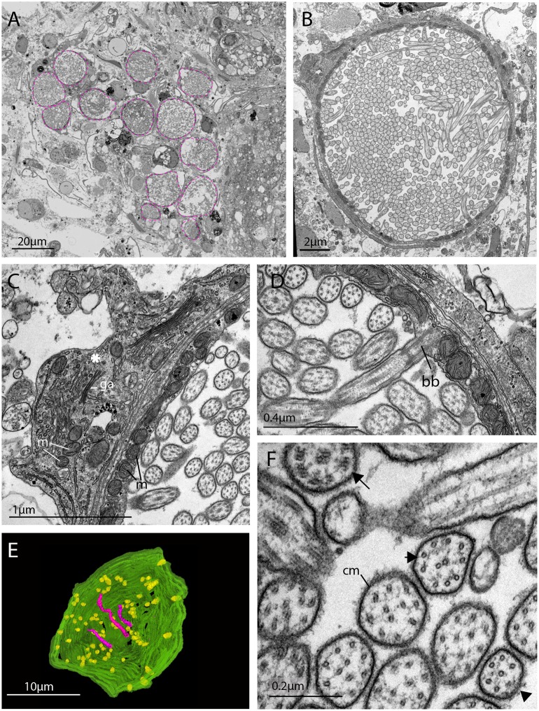
Figure 4.

Figure 4—video 1. Serial SEM images (101 × 500 nm sections = 50.5 μm total thickness) showing a cluster of ciliary phaosomes (intra-cellular vacuoles housing multiple cilia) that form the outer segment of the putative extraocular CPR cells in adult Maritigrella.
Download video file (13.1MB, mp4)
DOI: 10.7554/eLife.45465.012
This cluster is located to the left of the brain.
Figure 4—video 2. Serial SEM images (47 × 400 nm sections = 18.8 μm total thickness) showing a complete phaosome.
Download video file (5.9MB, mp4)
DOI: 10.7554/eLife.45465.013
At least 421 basal bodies were counted projecting cilia into the intra-cellular space that is formed by the cell itself. Note that in a nearby phaosome, the cilia appear to be arranged in a spirally coiled bundle.
Figure 4—video 3. Serial SEM images (72 x 100nm sections = 7.2μm total thickness) showing a third of a phaosome.
Download video file (10.9MB, mp4)
DOI: 10.7554/eLife.45465.014
Figure 4—video 4. A 3D reconstruction of series in Figure 4—video 3 shows that the cilia (pink) are unbranched and basal bodies (yellow) emerge all around the diameter of the cavity.
Download video file (2.2MB, mp4)
DOI: 10.7554/eLife.45465.015