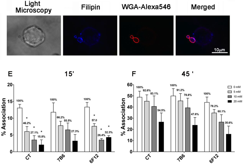
Figure 2 – Importance of sterols in the Hc association with macrophages and effect of m-β-CD treatment on the association with Hc.
(A) Light microscopy of Mφ-Hc interaction. (B and C) Filipin staining of cholesterol in J774.16 Mφ membranes and WGA-Alexa546 in Hc. (D) Merged images denoting the co-localization of filipin staining and WGA-Alexa546-Hc. Bar, 10 μm. (E) GFP-Hc yeast cells with or without opsonization with antibodies to HSP60 (mAb 7B6) or to M antigen (mAb 6F12) were incubated for 15 or (F) 45 minutes with J774.16 Mφs pre-treated with different concentrations of m-β-CD. Samples were analyzed by flow cytometry. Bars show the percentage of yeast-associated Mφs over the total number of Mφs analyzed. The numbers on the bars indicate the total infected Mφs in each system followed by the percentage determined in the setting of infection compared to controls (considered as 100%). The results shown are the average of three independent experiments. Each individual system showed statistical significance (P<0.0001) in comparison to its appropriate control.

