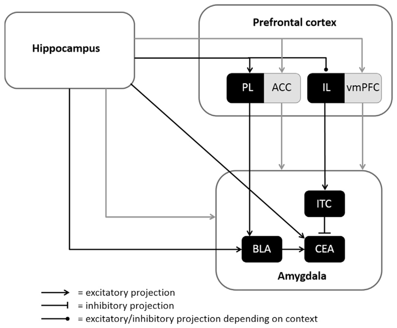
Fig. 1.
Neural networks of extinction. Neural networks for extinction have been investigated heavily in rodents and a network including at least the amygdala, hippocampus, and prefrontal areas has been identified. Substantial progress has been made in uncovering the subregions involved in extinction in rodents; the prelimibic area (PL) innervates the basolateral amygdala (BLA), which in turn projects to the central amygdala (CEA). The CEA controls conditioned responding and receives input from the infralimbic area (IL), mediated through the intercalated cells (ITC) of the amygdala. The hippocampus projects directly, and indirectly via the PL and IL, to the amygdala. In humans, it has been hypothesized that the same areas are involved in extinction learning. The anterior cingulate cortex (ACC) and the ventromedial prefrontal cortex (vmPFC) are assumed to constitute the human homologue of the PL and IL, respectively. Although the amygdala is generally assumed to be involved in fear learning and extinction in humans, neuroimaging evidence for this involvement is scarce. White areas constitute both the animal and human extinction network; black areas are specifically identified in animals; grey areas are related to the human extinction network.

