Fig. 3.

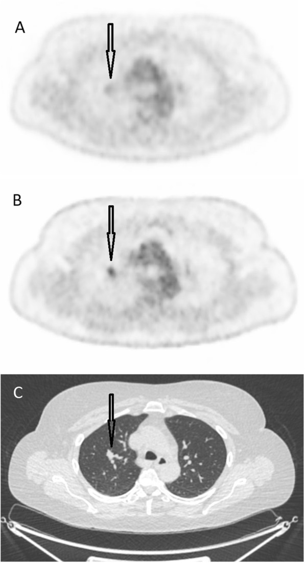
a) A transversal image from the Gemini TF. b) The same image from the Discovery MI and c) the corresponding CT image. The arrow indicates a tumour in the right lung, which was interpreted as malignant in b

a) A transversal image from the Gemini TF. b) The same image from the Discovery MI and c) the corresponding CT image. The arrow indicates a tumour in the right lung, which was interpreted as malignant in b