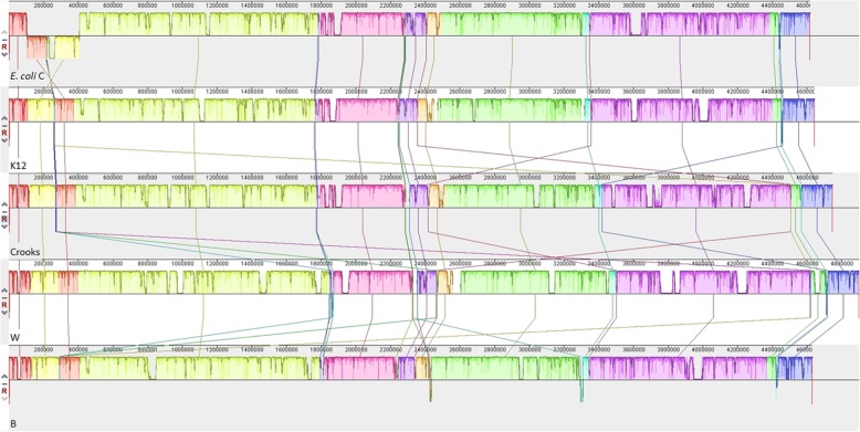
Fig. 5.
Genome alignment of five E. coli strains using Mauve. Each chromosome has been laid out horizontally and homologous blocks in each genome are shown as identically colored regions linked across genomes. The inverted region in E. coli C is shifted below the genome’s center axis. From the top: E. coli C, K12, Crooks, W, and B

